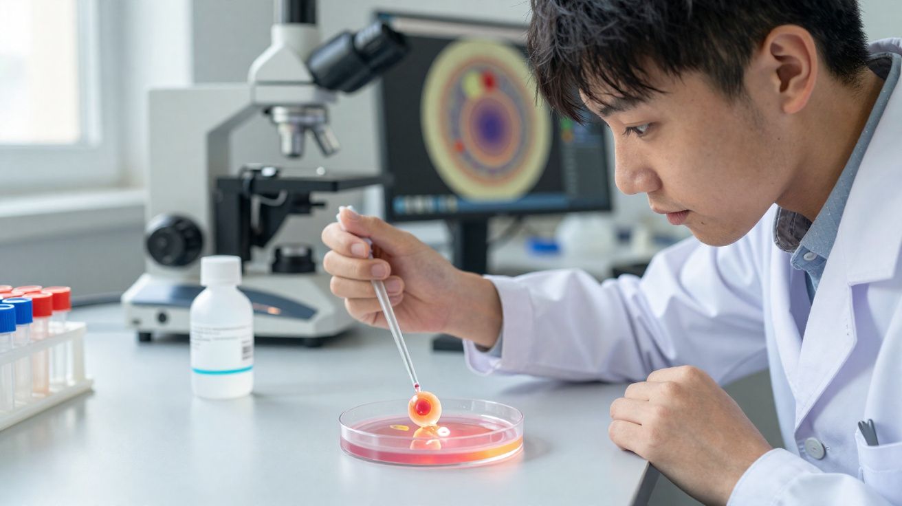
Scientists have created human embryo models from stem cells grown in the laboratory, offering an unprecedented look at the crucial first week after an embryo implants into the uterine wall.
Why the post-implantation stage matters for fertility and birth defects
A clearer understanding of what happens immediately after a fertilised egg embeds in the uterine lining could improve knowledge of fertility, early pregnancy loss, and developmental birth defects. However, ethical constraints and technical limitations have long restricted direct study of these pivotal early phases of human embryonic development.
"The drama is in the first month, the remaining eight months of pregnancy are mainly lots of growth," says molecular geneticist Jacob Hanna from the Weizmann Institute of Science in Israel.
"But that first month is still largely a black box. Our stem cell–derived human embryo model offers an ethical and accessible way of peering into this box."
Building human embryo models from genetically unmodified stem cells
An international team guided genetically unmodified, undifferentiated human-derived stem cells into intricate structures designed to mirror human embryonic development. The work highlights the striking self-organising capacity of human stem cells and builds on a recent breakthrough in producing embryo-like stem-cell systems, creating a new route to investigate processes that have previously been hidden by practical and ethical barriers.
Notably, these models include key features absent from earlier versions: the three lineages that give rise to the placenta and embryonic support tissues, alongside the cell layer that forms an embryo before it folds and develops into diverse tissues and organs.
Earlier studies found that stem cells taken from mouse embryos can still be steered to form both embryonic and supporting tissues, assembling into a structural stem-cell based embryo model (SEM) at the post-gastrulation stage, when embryonic cells organise into the three primary body tissue types.
"Here, we extend these findings to humans, while using only genetically unmodified human naïve embryonic stem cells," writes Hanna and colleagues.
"We proceeded to test the capacity to form embryo-like structures… that could mimic different stages of natural human in utero development."
What the human SEMs show up to 13–14 days post-fertilisation
The researchers identified optimal settings-such as cell numbers, proportions within mixed cell populations, and culture formulations-across multiple stages, beginning with implantation occurring 7–8 days after fertilisation.
"These human complete SEMs demonstrated developmental growth dynamics that resemble key hallmarks of post-implantation stage embryogenesis up to 13-14 days post-fertilization," the authors write.
These models depict the formation of all recognised early embryo lineages and components, including the epiblast, hypoblast, extraembryonic mesoderm, trophoblast, and yolk sac. When compared with a reference dataset, cell profiles from the human SEMs dataset closely matched gene-expression patterns and cell-type composition seen in human embryos shortly after implantation.
The authors emphasise that human SEMs are not the same as embryos, but they provide a model with major research potential.
"Many failures of pregnancy occur in the first few weeks, often before the woman even knows she's pregnant. That's also when many birth defects originate, even though they tend to be discovered much later," Hanna says.
"Our models can be used to reveal the biochemical and mechanical signals that ensure proper development at this early stage, and the ways in which that development can go wrong."
The study has been published in Nature.


Comments
No comments yet. Be the first to comment!
Leave a Comment