Far from the core of a tumour, quiet molecular messages are already altering distant organs, discreetly laying the groundwork for cancer’s next step.
Scientists are now following those messages at the nanoscale and recreating them in the laboratory, with the goal of turning one of cancer’s hidden advantages into a highly precise way to fight it.
Cancer’s secret bubbles that prepare metastasis
Most deaths from cancer are caused not by the primary tumour itself, but by metastasis-secondary tumours that emerge in organs a long way from where the disease began. For a long time, this spread looked largely chance-driven, as though cells simply detached and became trapped wherever the circulation carried them.
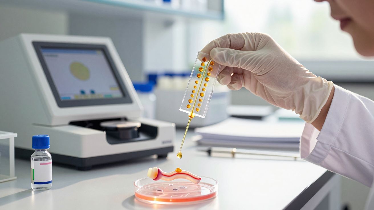
That explanation is being rapidly replaced. Tumours do more than release wandering cancer cells: they also shed vast numbers of nanoscale lipid-coated “bubbles” called extracellular vesicles (EVs). These particles transport proteins, genetic material and fats through the blood and other bodily fluids, functioning like miniature parcels that deliver instructions to distant tissues.
Before cancer cells show up, EVs can already be remodelling the destination organ, forming what researchers describe as a pre-metastatic niche.
Research groups associated with the McGill University Health Centre Research Institute have demonstrated how EVs can “prime” otherwise healthy organs. They can make blood vessel walls more permeable, recruit immune cells that tolerate tumours rather than attacking them, and encourage the formation of new blood vessels that may later nourish arriving cancer cells.
In mouse models, the effect is striking: injecting tumour-derived vesicles alone can kick-start these early organ changes even when no cancer cells are present. Metastasis looks less like a roll of the dice and more like a coordinated strategy-prepare the ground first, then send in the cells.
How extracellular vesicles (EVs) bend cell behaviour
When EVs reach a target tissue, they can attach to cells or be taken up by them. Once inside, their cargo can reprogramme cellular machinery. Some EVs deliver RNA fragments that switch genes on or off; others carry proteins that change how cells adhere to their surroundings or how strongly they react to inflammation.
These shifts can nudge normal cells into tumour-supporting roles. Cells lining blood vessels may become more permissive, allowing circulating tumour cells to pass through more readily. Immune cells can pivot from attack to repair-a response that, in this setting, benefits the tumour. Fibroblasts, which help form the tissue’s structural framework, may be driven to produce a matrix that better supports cancer growth.
Metastasis can begin well before a scan detects a mass; the dialogue between tumour and organ often starts with these nanoscale messengers.
Also in the feed (unrelated headlines):
Building artificial bubbles to decode cancer’s messages
A key obstacle in EV research is practical rather than conceptual: natural vesicles are inherently inconsistent. Even EVs released by a single tumour can differ widely in size, composition and behaviour. Their properties can also shift with time, treatment, and even the way samples are collected. That variability makes tightly controlled experiments difficult.
To get around this, a team led by Julia V. Burnier at McGill has leaned on biomimicry. They manufacture artificial vesicles known as liposomes. Liposomes imitate the basic architecture of EVs but can be produced with far greater uniformity and tunability. Using microfluidic mixers, researchers can precisely adjust features such as particle size, surface charge and lipid composition.
Size and charge matter more than you think
Experiments with these engineered particles show how much “physics” influences biology. When 100-nanometre liposomes were compared with 300-nanometre versions, the smaller particles entered certain cell types far more efficiently. This implies tumours may favour particular size ranges when sending their molecular “post” around the body.
Surface charge proved equally influential. Liposomes engineered with a negative zeta potential of roughly -40 millivolts were taken up more readily by human endothelial cells (the cells that line blood vessels). Altering the charge changed how many particles cells internalised.
Simply tweaking size or charge can increase-or reduce-how readily cells engulf these nanobubbles.
By modifying lipid composition, the team could reproduce the distinctive “signature” of vesicles released by specific cancers. That, in turn, made it possible to test how those signatures affect adhesion, cell signalling pathways and immune responses. Because liposomes are synthetic, batches can be replicated closely, helping researchers isolate cause and effect far more cleanly than with natural EVs.
- Size influences how easily liposomes enter different cell types.
- Surface charge shapes interactions with cell membranes and blood proteins.
- Lipid composition affects immune recognition and downstream signalling.
A related piece of the puzzle: standardising EV measurements
A closely connected challenge-beyond simply producing uniform liposomes-is agreeing on how EVs themselves should be measured and compared between laboratories. Differences in isolation methods, storage conditions and readouts can change what is labelled as an EV “signal”. As the field advances, standardised reporting frameworks and cross-lab benchmarking will be essential for translating discoveries into robust diagnostics and therapies.
In practice, teams often combine multiple tools-such as particle tracking approaches, high-resolution microscopy and molecular profiling-to ensure that the vesicles being studied are correctly characterised. The more consistent these methods become, the easier it is to identify which EV features truly matter for metastasis and which are artefacts of handling.
Turning cancer’s trick into a treatment tool
Once liposomes can be engineered to behave like tumour vesicles, an obvious next step follows: use them against the very disease they emulate. Instead of transporting pro-metastatic instructions, these synthetic bubbles can deliver drugs-or act as decoys that interfere with harmful signalling.
Work reported in journals including Science Advances indicates that bio-inspired liposomes can carry chemotherapy agents directly into tumour cells. Because their surfaces resemble natural EVs, cancer cells-often highly active in endocytosis-may take them up readily.
Targeted drug delivery with fewer side effects
In one study, liposomes packed with doxorubicin (a widely used chemotherapy) were tested in glioblastoma cell cultures. Compared with the free drug, the liposome carriers killed cancer cells more effectively. They also did less collateral damage to neighbouring healthy cells, in part by limiting how much drug leaked into the surrounding environment.
The aim is to conceal toxic drugs inside “camouflaged” bubbles-hitting tumours harder while reducing harm to the rest of the body.
Another approach avoids drug payloads altogether. Empty liposomes can serve as competitors: when these “decoy” bubbles circulate, they can occupy receptors or binding sites that tumour EVs would typically use. Early evidence suggests this may reduce how many malignant vesicles reach their preferred target cells, weakening the formation of pre-metastatic niches.
Taken together, liposomes could play a double part-both as targeted delivery vehicles and as blockers of damaging cell-to-cell communication. This shifts attention from shrinking the primary tumour alone to disrupting the wider messaging network that enables cancer to spread and adapt.
Hurdles on the way to treating patients
Moving these nano-strategies from Petri dish to clinic raises several difficult questions. A major one is specificity: a liposome circulating in the bloodstream will encounter many cell types, and it must preferentially engage cancer cells rather than healthy tissues.
To improve targeting, researchers attach ligands-small molecules or antibodies-that bind receptors found at high levels on tumour cells. However, these markers can vary between cancers (breast, lung, melanoma and others) and can change as tumours evolve, meaning each disease may require a different liposome design.
Survival in the bloodstream is another challenge. Circulating particles face enzymes, immune surveillance, and filtration by organs such as the liver, all of which can clear liposomes quickly. Coating particles with polyethylene glycol (PEG) can prolong circulation time, but PEG itself may provoke immune responses in some patients.
Manufacturing is a further hurdle. Scaling up microfluidic production while keeping batches truly consistent is technically demanding. Particle size distributions, sterility testing and stability assessments must all meet stringent pharmaceutical standards before clinical trials can advance beyond the earliest stages.
Beyond the lab: safety, regulation and access
Even if the engineering challenges are solved, nano-medicines still have to clear real-world barriers: regulators will expect clear evidence on biodistribution, long-term safety, immune effects and interactions with other medicines. Health systems will also need manufacturing processes that are reliable and affordable at scale, otherwise promising liposome-based treatments may remain confined to specialist centres.
Equally important is ensuring that diagnostic EV tests and liposome therapies, if validated, are deployed in a way that improves outcomes broadly-rather than widening gaps in access between different regions and patient groups.
What “nanobubbles” and “liposomes” really are
The terminology can feel opaque, so it helps to define the basics. Extracellular vesicles are natural packages released by cells. Each is enclosed by a lipid membrane-a double layer of fat molecules similar to the cell’s outer membrane. Inside, EVs may contain DNA fragments, RNA, proteins and lipids.
Liposomes share the same core structure but are made artificially. Researchers mix defined lipids under tightly controlled conditions, commonly using microfluidic systems that drive fluids through tiny channels. By changing the recipe and process parameters, they create spheres with predictable size and composition, sometimes trapping water-soluble drug molecules inside.
| Feature | Natural extracellular vesicles | Synthetic liposomes |
|---|---|---|
| Origin | Released by living cells | Produced in microfluidic or chemical systems |
| Cargo | Proteins, RNA, lipids, DNA fragments | Selected drugs or experimental molecules |
| Control | Highly variable | Precisely adjustable |
| Main use in research | Biomarkers and signalling research | Drug delivery and mechanistic experiments |
How this might affect patients in the future
If these technologies reach maturity, they could reshape cancer care at multiple points. In diagnosis, blood tests that profile EVs may indicate whether a tumour is already establishing new niches, even when imaging still appears clear. That could justify earlier or more intensive treatment for patients at high risk of metastasis.
In treatment, clinicians might pair standard chemotherapy with liposome-based formulations. One plausible model is using a lower systemic dose alongside nano-carriers that concentrate additional drug within the tumour. Another is deploying decoy liposomes between treatment cycles to reduce metastatic signalling while the patient recovers.
There are also genuine risks. Any new nano-medicine prompts questions about long-term accumulation, unforeseen immune reactions and interactions with other therapies. Carefully designed clinical trials, extended follow-up and transparent reporting will be needed to determine who benefits-and who might be harmed.
Even if liposome therapies take time to reach routine practice, the central insight is already altering how oncologists think about disease spread: metastasis is not merely a late-stage accident, but a process that can be orchestrated in advance by billions of invisible bubbles. Mapping that choreography opens up new opportunities-from earlier detection to more targeted, better-tolerated treatments.


Comments
No comments yet. Be the first to comment!
Leave a Comment